File:Gingival sulcus.PNG
Jump to navigation
Jump to search
No higher resolution available.
Gingival_sulcus.PNG (221 × 296 pixels, file size: 59 KB, MIME type: image/png)
Captions
Captions
Add a one-line explanation of what this file represents
Summary[edit]
| Description |
Deutsch: Zwischen der Zahnwurzel und dem Alveolarknochen sind deutlich die Sharpey-Faser-Bündel zu erkennen.
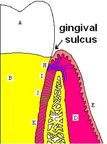
English: Gingival and periodontal pockets are extensions of the gingival sulcus, which exists in health. A, crown of the tooth, covered by enamel. B, root of the tooth, covered by cementum. C, alveolar bone. D, subepithelial connective tissue. E, oral epithelium. H, principle gingival fibers. I, alveolar crest fibers of the PDL. J, horizontal fibers of the PDL. K, oblique fibers of the PDL.
Español: Surco gingival.
Suomi: Parodontium-kudoksia. |
| Date | |
| Source | Image:The Periodontium.jpg |
| Author | User:Mikael Häggström |
Licensing[edit]
| I, the copyright holder of this work, release this work into the public domain. This applies worldwide. In some countries this may not be legally possible; if so: I grant anyone the right to use this work for any purpose, without any conditions, unless such conditions are required by law. |
File history
Click on a date/time to view the file as it appeared at that time.
| Date/Time | Thumbnail | Dimensions | User | Comment | |
|---|---|---|---|---|---|
| current | 18:05, 21 October 2007 | | 221 × 296 (59 KB) | Mikael Häggström (talk | contribs) | {{Information |Description= |Source= Image:The Periodontium.jpg |Date= 2007-10-21 |Author= User:Mikael Häggström }} |
You cannot overwrite this file.
File usage on Commons
There are no pages that use this file.
File usage on other wikis
The following other wikis use this file:
- Usage on ar.wikipedia.org
- Usage on bn.wikipedia.org
- Usage on de.wikipedia.org
- Usage on en.wikipedia.org
- Usage on es.wikipedia.org
- Usage on fa.wikipedia.org
- Usage on fi.wikipedia.org
- Usage on fr.wikipedia.org
- Usage on fr.wiktionary.org
- Usage on nl.wikipedia.org
- Usage on pt.wikipedia.org
- Usage on ro.wikipedia.org