File:FluorescentCells.jpg
Jump to navigation
Jump to search
No higher resolution available.
FluorescentCells.jpg (512 × 512 pixels, file size: 56 KB, MIME type: image/jpeg)
Captions
Captions
Add a one-line explanation of what this file represents
Summary[edit]
| Description |
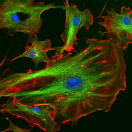
English: a
This image is made from a Molecular Probes demo slide:
Deutsch: Endothelzellen aus der Inneren Wand (Endothel) von Lungenarterien des Rindes unter dem Mikroskop. Die Zellkerne sind mit DAPI blau markiert. Die Mikrotubuli wurden über einen Antikörper grün markiert. Mit rot fluoreszierendem Phalloidin wurden die Aktinfilamente markiert.
Français : Cellulles endothéliales vues au microscope. En bleu, noyaux marqués au DAPI. En vert, microtubules marqués par un anticorps. En rouge, actine marquée à la phalloïdine.
Magyar: Fluoreszcenciamikroszkópos felvétel marha tüdőartéria endotélsejtjeiről (Molecular Probes FluoCells prepared slide #2 (F14781)). A sejtmagok DAPI-val vannak festve (kék), a mikrotubulusokhoz anti-α-tubulin egéranitest, ahhoz pedig BODIPY FL-el jelölt anti-egér kecske-IgG van kapcsolva (zöld), míg az aktin filamentumok Texas Red-X-el kapcsolt falloidinnal vannak jelölve (vörös). A kép három felvétel szuperpozíciójával készült. Hamis színek.
Lietuvių: Citoskeletas. Aktino filamentai – raudona, mikrovamzdeliai – žalia, branduolys – mėlyna spalva.
Română: Sub microscop Celule endoteliale . microtubulii sunt de culoare verde, iar filamentele de actină sunt roşii, pe când nucleul celulei este colorat albastru
Русский: Цитоскелет эукариот. Актиновые микрофиламенты окрашены в красный (фаллоидином, связанным с TRITC), микротрубочки — в зеленый (антителами, связанными с FITC), ядра клеток — в голубой цвет (DAPI). Клетки эндотелия лёгочной артерии быка.
Українська: Цитоскелет еукаріот. Актинові мікрофіламенти забарвлені в червоний колір, мікротрубочки — в зелений, ядра кліток — в блакитний |
| Source | http://rsb.info.nih.gov/ij/images/ |
| Author | |
| Permission (Reusing this file) |
example image from the ImageJ-Programmpaket (public domain) |
Original file[edit]
This image has been taken from the German Wikipedia
The original uploader is de:Benutzer:Jan R. The original upload was at 4th December 2005.
Original description[edit]
This image is made from a Molecular Probes demo slide:
Cells: bovine pulmonary arthery endothelial cells
Blue: nucleus stained with DAPI
Green: Tubulin stained with Bodipy FL goat anti-mouse IgG
Red: F-Actin stained with Texas Red X-Phalloidin
(description from [1])
Quelle: Beispielsbild aus dem ImageJ-Programmpaket (public domain), siehe http://rsb.info.nih.gov/ij/
Licensing[edit]
This work is in the public domain in the United States because it is a work prepared by an officer or employee of the United States Government as part of that person’s official duties under the terms of Title 17, Chapter 1, Section 105 of the US Code.
Note: This only applies to original works of the Federal Government and not to the work of any individual U.S. state, territory, commonwealth, county, municipality, or any other subdivision. This template also does not apply to postage stamp designs published by the United States Postal Service since 1978. (See § 313.6(C)(1) of Compendium of U.S. Copyright Office Practices). It also does not apply to certain US coins; see The US Mint Terms of Use.
|
 | |
| This file has been identified as being free of known restrictions under copyright law, including all related and neighboring rights. | ||
File history
Click on a date/time to view the file as it appeared at that time.
| Date/Time | Thumbnail | Dimensions | User | Comment | |
|---|---|---|---|---|---|
| current | 15:07, 24 March 2006 | | 512 × 512 (56 KB) | Splette (talk | contribs) | {{Information |Description = Endothelial cells under the microscope. Nuclei are stained blue with DAPI, microtubles are marked green by an antibody and actin filaments are labelled red with phalloidin. |Source = http://rsb.info.nih.gov/ij |Date = |Author |
You cannot overwrite this file.
File usage on Commons
The following 5 pages use this file:
File usage on other wikis
The following other wikis use this file:
- Usage on af.wikipedia.org
- Usage on ar.wikipedia.org
- Usage on ast.wikipedia.org
- Usage on az.wikipedia.org
- Usage on be.wikipedia.org
- Usage on bg.wikipedia.org
- Usage on bn.wikipedia.org
- Usage on bs.wikipedia.org
- Usage on ca.wikipedia.org
- Usage on ckb.wikipedia.org
- Usage on cs.wikipedia.org
- Usage on cy.wikipedia.org
- Usage on da.wikipedia.org
- Usage on de.wikipedia.org
- Ultraviolettstrahlung
- Mikrotubulus
- Skelett
- Cytoskelett
- Aktin
- 4′,6-Diamidin-2-phenylindol
- Fluoreszenzmikroskopie
- Listeriose
- Fluoreszenzmarkierung
- Wikipedia Diskussion:Hauptseite/Artikel des Tages/Archiv/Vorschläge/2018/Q3
- Wikipedia:Hauptseite/Archiv/5. August 2018
- Wikipedia Diskussion:Hauptseite/Artikel des Tages/Archiv/Vorschläge/2019/Q1
- Wikipedia:Hauptseite/Archiv/23. März 2019
- Usage on de.wikibooks.org
- Usage on de.wikiversity.org
- Usage on en.wikipedia.org
View more global usage of this file.