File:Clinical lectures on stricture of the urethra and enlargement of the prostate (1901) (14783994362).jpg
From Wikimedia Commons, the free media repository
Jump to navigation Jump to search

Original file(1,608 × 632 pixels, file size: 63 KB, MIME type: image/jpeg)

Captions

Captions

Add a one-line explanation of what this file represents

Summary[edit]


Description
English:

Identifier: clinicallectures1901frey (find matches)
Title: Clinical lectures on stricture of the urethra and enlargement of the prostate
Year: 1901 (1900s)
Authors: Freyer, P. J. (Peter Johnston), Sir, 1851-1921
Subjects: Prostate Prostate Urethra Urethra Urethra Prostate
Publisher: New York : William Wood & Co.
Contributing Library: Francis A. Countway Library of Medicine
Digitizing Sponsor: Open Knowledge Commons and Harvard Medical School

View Book Page: Book Viewer
About This Book: Catalog Entry
View All Images: All Images From Book
Click here to view book online to see this illustration in context in a browseable online version of this book.

Text Appearing Before Image:
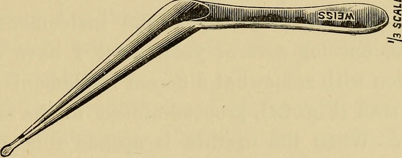
Fig. 22. edges of the wound are held aside by catch-forceps, or by aloop of silk passed on either side through the margins of theurethra. The beak of the staff is then turned round andhooks up the upper end of the incision. All bleeding havingbeen arrested and the wound carefully sponged, the face ofthe stricture comes into view, and the opening is searched for,
Text Appearing After Image:
Fig. 23. and when found a small probe-shaped director (Fig. 22) ispushed through into the bladder. The stricture is cut on itslower aspect by passing along the director a long narrowknife. The beak of a small blunt gorget (Fig. 23) is insertedin the groove of the director and guided thus into the bladder.The staff, forceps, and director are removed, and a stoutflexible catheter, No. 12 English scale, introduced through the 60 OPERATIVE TREATMENT OF STRICTURE urethra and along the gorget into the bladder, in whichposition it is fixed. Over this the edges of the wound arepartially closed by silkworm sutures, and the dressingsapplied. The catheter is retained for three or four days, or longerif it does not cause irritation, the urine flowing thereby.After its removal a full - sized steel dilator is introducedevery second or third day whilst the wound is granulating,and at more distant intervals when it completely closes. This is a sound operation. It is sometimes a difficult one,requirin

Note About Images

Please note that these images are extracted from scanned page images that may have been digitally enhanced for readability - coloration and appearance of these illustrations may not perfectly resemble the original work.
Date
Source

https://www.flickr.com/photos/internetarchivebookimages/14783994362/

Author Internet Archive Book Images
Permission
(Reusing this file)
At the time of upload, the image license was automatically confirmed using the Flickr API. For more information see Flickr API detail.
Flickr tags
InfoField
  • bookid:clinicallectures1901frey
  • bookyear:1901
  • bookdecade:1900
  • bookcentury:1900
  • bookauthor:Freyer__P__J___Peter_Johnston___Sir__1851_1921
  • booksubject:Prostate
  • booksubject:Urethra
  • bookpublisher:New_York___William_Wood___Co_
  • bookcontributor:Francis_A__Countway_Library_of_Medicine
  • booksponsor:Open_Knowledge_Commons_and_Harvard_Medical_School
  • bookleafnumber:64
  • bookcollection:medicalheritagelibrary
  • bookcollection:francisacountwaylibrary
  • bookcollection:americana
Flickr posted date
InfoField
30 July 2014

Licensing[edit]

This image was taken from Flickr's The Commons. The uploading organization may have various reasons for determining that no known copyright restrictions exist, such as:
  1. The copyright is in the public domain because it has expired;
  2. The copyright was injected into the public domain for other reasons, such as failure to adhere to required formalities or conditions;
  3. The institution owns the copyright but is not interested in exercising control; or
  4. The institution has legal rights sufficient to authorize others to use the work without restrictions.

More information can be found at https://flickr.com/commons/usage/.


Please add additional copyright tags to this image if more specific information about copyright status can be determined. See Commons:Licensing for more information.
This image was originally posted to Flickr by Internet Archive Book Images at https://flickr.com/photos/126377022@N07/14783994362. It was reviewed on 17 September 2015 by FlickreviewR and was confirmed to be licensed under the terms of the No known copyright restrictions.

17 September 2015

File history

Click on a date/time to view the file as it appeared at that time.

Date/TimeThumbnailDimensionsUserComment
current14:52, 17 September 2015Thumbnail for version as of 14:52, 17 September 20151,608 × 632 (63 KB) (talk | contribs)== {{int:filedesc}} == {{subst:chc}} {{information |description={{en|1=<br> '''Identifier''': clinicallectures1901frey ([https://commons.wikimedia.org/w/index.php?title=Special%3ASearch&profile=default&fulltext=Search&search=insource%3A%2Fclinicallectu...

There are no pages that use this file.