File:Chordoma3.JPG
Jump to navigation
Jump to search
Size of this preview: 652 × 599 pixels. Other resolutions: 261 × 240 pixels | 522 × 480 pixels | 695 × 639 pixels.
Original file (695 × 639 pixels, file size: 41 KB, MIME type: image/jpeg)
Captions
Captions
Add a one-line explanation of what this file represents
Summary[edit]
More images of this case:  
|
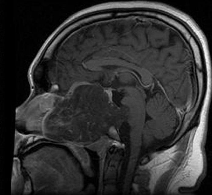
| Description | MRI of extensive clival chordoma in 17-year old male patient, sagittal view. Tumor in the nasopharynx extending from nasal cavity to brainstem posteriorly is clearly visible. |
| Date | |
| Source | Hassan S, Abdullah JM, Wan Din SJ, Idris Z. Combined use of maxillomandibular swing approach and neurosurgical ultrasonic aspirator in the management of extensive clival chordoma: a case report. Journal of Medical Case Reports. 2, 49. 2008. doi:10.1186/1752-1947-2-49. PMID 18279530. |
| Author | Hassan S, Abdullah JM, Wan Din SJ, Idris Z |
| Permission (Reusing this file) |
BioMedCentral licence |
Licensing[edit]
This file is licensed under the Creative Commons Attribution 2.0 Generic license.
- You are free:
- to share – to copy, distribute and transmit the work
- to remix – to adapt the work
- Under the following conditions:
- attribution – You must give appropriate credit, provide a link to the license, and indicate if changes were made. You may do so in any reasonable manner, but not in any way that suggests the licensor endorses you or your use.
File history
Click on a date/time to view the file as it appeared at that time.
| Date/Time | Thumbnail | Dimensions | User | Comment | |
|---|---|---|---|---|---|
| current | 00:19, 23 February 2008 | | 695 × 639 (41 KB) | Filip em (talk | contribs) | {{Information |Description=MRI of extensive clival chordoma in 17-year old male patient, axial view. Tumor in the nasopharynx extending from nasal cavity to brainstem posteriorly is clearly visible. |Source=Hassan S, Abdullah JM, Wan Din SJ, Idris Z. Co |
You cannot overwrite this file.
File usage on Commons
The following 3 pages use this file:
File usage on other wikis
The following other wikis use this file:
- Usage on ar.wikipedia.org
- Usage on bs.wikipedia.org
- Usage on en.wikipedia.org
- Usage on en.wikibooks.org
- Usage on ko.wikipedia.org
- Usage on outreach.wikimedia.org
- Usage on ta.wikipedia.org