File:Cervical Xray Extension view.jpg
Jump to navigation
Jump to search

Size of this preview: 422 × 599 pixels. Other resolutions: 169 × 240 pixels | 338 × 480 pixels | 541 × 768 pixels | 721 × 1,024 pixels | 1,411 × 2,003 pixels.
Original file (1,411 × 2,003 pixels, file size: 138 KB, MIME type: image/jpeg)
Captions
Captions
Add a one-line explanation of what this file represents
Summary[edit]
| Description |
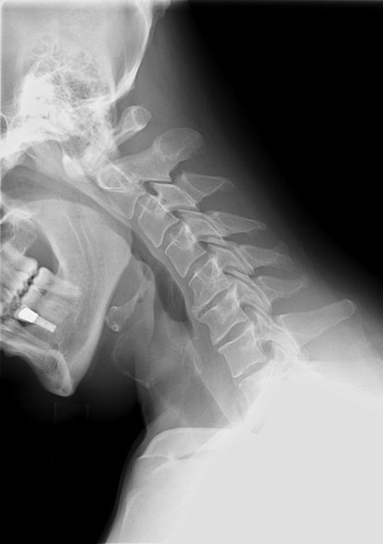
English: X-ray of cervical spine (neck) in flexion (bending forward). This series of x-rays were part of pre-surgical evaluation to help identify spinal instability. Patient is a 37 year old male with a history of multiple neck traumas with pain and muscle spasms and dental implant in lower jaw. Excerpt from radiologist's report:
Français : Ragioagraphie aux raysons X du rachis cervical (cou) en flexion. Cette série de radiographies faisaient partie de l'évaluation pré-chirurgicale pour aider à identifier une instabilité vertébrale. Le patient est un homme de 37 ans ayant des antécédents de traumatismes multiples cou avec des spasmes et des douleurs musculaires et implant dentaire à la mâchoire inférieure. Extrait du rapport du radiologiste:
|
| Date | |
| Source | own medical image, work for hire |
| Author | Stillwaterising |
| Other versions |
Derivative works of this file: |
Magnification 0.4x, converted from lossy DICOM file
Licensing[edit]
I, the copyright holder of this work, hereby publish it under the following license:
| This file is made available under the Creative Commons CC0 1.0 Universal Public Domain Dedication. | |
| The person who associated a work with this deed has dedicated the work to the public domain by waiving all of their rights to the work worldwide under copyright law, including all related and neighboring rights, to the extent allowed by law. You can copy, modify, distribute and perform the work, even for commercial purposes, all without asking permission.
|
File history
Click on a date/time to view the file as it appeared at that time.
| Date/Time | Thumbnail | Dimensions | User | Comment | |
|---|---|---|---|---|---|
| current | 21:51, 10 November 2010 | | 1,411 × 2,003 (138 KB) | Stillwaterising (talk | contribs) | crop and cleanup |
| 21:19, 10 November 2010 |  | 1,668 × 2,008 (126 KB) | Stillwaterising (talk | contribs) | {{Information |Description={{en|1=xray}} |Source={{own}} |Author=Stillwaterising |Date= |Permission= |other_versions= }} |
You cannot overwrite this file.
File usage on Commons
The following 3 pages use this file:
File usage on other wikis
The following other wikis use this file:
- Usage on ca.wikipedia.org