File:Cerebroventricular system.png
Jump to navigation
Jump to search

Size of this preview: 800 × 363 pixels. Other resolutions: 320 × 145 pixels | 640 × 290 pixels | 1,024 × 464 pixels | 2,137 × 969 pixels.
Original file (2,137 × 969 pixels, file size: 662 KB, MIME type: image/png)
Captions
Captions
Add a one-line explanation of what this file represents
Summary[edit]
| Description |
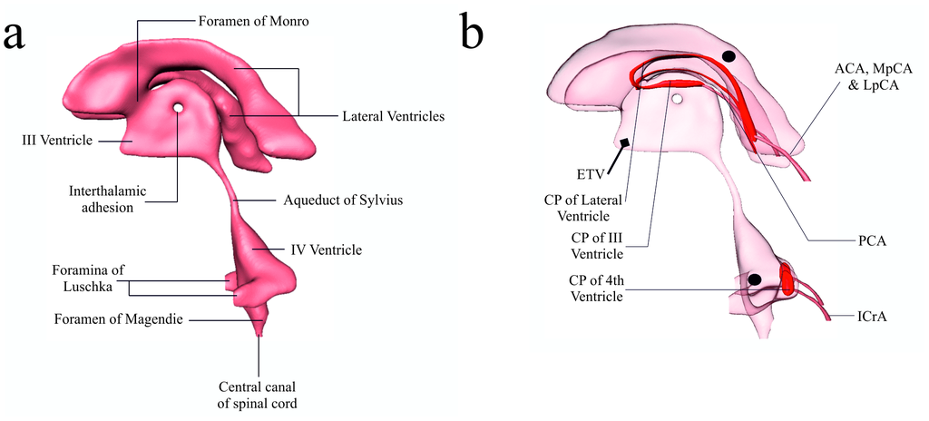
English: The cerebroventricular system, along with the choroid plexuses and some feeding arteries.
(a) CSF circulates through the four brain ventricles and in the subarachnoid space surrounding the brain and spinal cord. (b) View of the ventricular system along with the choroid plexus (CP) of the lateral (LV), 3rd and 4th ventricle. The arteries supplying these plexuses (not all are presented in the figure) are the anterior and lateral posterior choroidal arteries (ACA & LpCA), the P2 segment of the posterior cerebellar artery (PCA) and the medial posterior choroidal artery (MpCA) [12]. The MpCA arises from the PCA (or its branches) and enters the roof of the third ventricle [74] which feeds the CP situated there. Finally, the CP of the fourth ventricle is supplied by the posterior inferior cerebellar artery (PICA), inferior cerebellar artery (ICA) and superior cerebellar artery (SCA) [75]. ETV is the location of perforation of the floor of the third ventricle during endoscopic third ventriculostomy. [•] represents the locations of measurement for the deduction of the pressure difference between the lateral and fourth ventricles. |
| Date | Published: December 31, 2013 |
| Source | Vardakis JC, Tully BJ, Ventikos Y (2013) Exploring the Efficacy of Endoscopic Ventriculostomy for Hydrocephalus Treatment via a Multicompartmental Poroelastic Model of CSF Transport: A Computational Perspective. PLoS ONE 8(12): e84577. https://doi.org/10.1371/journal.pone.0084577 |
| Author | John C. Vardakis, Brett J. Tully, Yiannis Ventikos |
Licensing[edit]
This file is licensed under the Creative Commons Attribution 4.0 International license.
- You are free:
- to share – to copy, distribute and transmit the work
- to remix – to adapt the work
- Under the following conditions:
- attribution – You must give appropriate credit, provide a link to the license, and indicate if changes were made. You may do so in any reasonable manner, but not in any way that suggests the licensor endorses you or your use.
File history
Click on a date/time to view the file as it appeared at that time.
| Date/Time | Thumbnail | Dimensions | User | Comment | |
|---|---|---|---|---|---|
| current | 07:55, 9 June 2019 | | 2,137 × 969 (662 KB) | Was a bee (talk | contribs) | {{Information |Description={{en|1=The cerebroventricular system, along with the choroid plexuses and some feeding arteries. (a) CSF circulates through the four brain ventricles and in the subarachnoid space surrounding the brain and spinal cord. (b) View of the ventricular system along with the choroid plexus (CP) of the lateral (LV), 3rd and 4th ventricle. The arteries supplying these plexuses (not all are presented in the figure) are the anterior and lateral posterior choroidal arteries (A... |
You cannot overwrite this file.
File usage on Commons
There are no pages that use this file.