File:Basal ganglia in Parkinson's disease.png
Jump to navigation
Jump to search

Size of this preview: 608 × 599 pixels. Other resolutions: 243 × 240 pixels | 487 × 480 pixels | 779 × 768 pixels | 1,061 × 1,046 pixels.
Original file (1,061 × 1,046 pixels, file size: 461 KB, MIME type: image/png)
Captions
Captions
Add a one-line explanation of what this file represents
Summary[edit]
| Description |
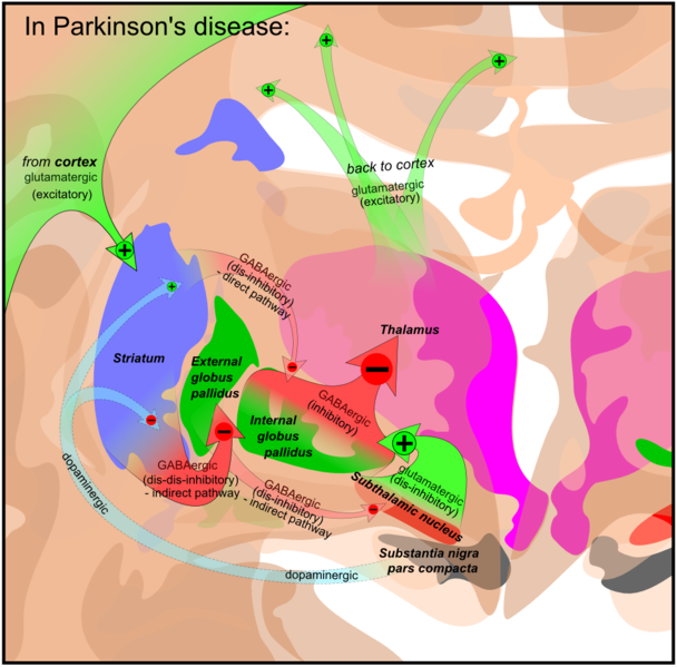
English: Circuits of the basal ganglia in Parkinson's disease. Picture shows 2 coronal slices have been superimposed to include the involved basal ganglia. Green arrows refer to excitatory (+) glutamatergic pathways, red arrows refer to inhibitory (-) GABAergic pathways and turquoise arrows refer to dopaminergic pathways, that are excitatory on the direct pathway and inhibitory on the indirect pathway. Note that dis-inhibitory pathways in effect are excitatory on the feedback to the cortex, while dis-dis-inhibitory pathways in effect are inhibitory. |
| Date | |
| Source | Derivative of File:Basal ganglia circuits.png |
| Author | Mikael Häggström, based on image by Andrew Gillies/User:Anaru |

|
File:Basal ganglia in Parkinson's disease.svg is a vector version of this file. It should be used in place of this PNG file when not inferior.
File:Basal ganglia in Parkinson's disease.png → File:Basal ganglia in Parkinson's disease.svg
For more information, see Help:SVG. |
 |
Basal ganglia gallery[edit]
-

-

-

-
 Basal ganglia without Parkinson's disease.png.
(Derived from Basal ganglia circuits.svg with small change)


Licensing[edit]
I, the copyright holder of this work, hereby publish it under the following license:
This file is licensed under the Creative Commons Attribution-Share Alike 3.0 Unported license.
- You are free:
- to share – to copy, distribute and transmit the work
- to remix – to adapt the work
- Under the following conditions:
- attribution – You must give appropriate credit, provide a link to the license, and indicate if changes were made. You may do so in any reasonable manner, but not in any way that suggests the licensor endorses you or your use.
- share alike – If you remix, transform, or build upon the material, you must distribute your contributions under the same or compatible license as the original.
File history
Click on a date/time to view the file as it appeared at that time.
| Date/Time | Thumbnail | Dimensions | User | Comment | |
|---|---|---|---|---|---|
| current | 07:34, 25 August 2010 | | 1,061 × 1,046 (461 KB) | Mikael Häggström (talk | contribs) | Removed pars reticularis pathways for simplification, as they appear to be of relatively minor importance in Parkinson's disease. |
| 10:29, 29 May 2010 |  | 1,061 × 1,046 (494 KB) | Mikael Häggström (talk | contribs) | Simplified text colors. Smaller arrow from cortex. Easier to see text over SN. | |
| 10:24, 22 May 2010 |  | 1,061 × 1,046 (505 KB) | Mikael Häggström (talk | contribs) | Text adjustment. Glutaminergic --> Glutamatergic | |
| 14:37, 11 May 2010 |  | 1,061 × 1,046 (508 KB) | Mikael Häggström (talk | contribs) | reinserted nigra- thalamus | |
| 13:58, 11 May 2010 |  | 1,061 × 1,046 (497 KB) | Mikael Häggström (talk | contribs) | {{Information |Description={{en|1=f}} |Source={{own}} |Author=Mikael Häggström |Date= |Permission= |other_versions= }} |
You cannot overwrite this file.
File usage on Commons
The following 10 pages use this file:
- File:Basal ganglia circuits.png
- File:Basal ganglia circuits.svg
- File:Basal ganglia circuits zh-hans.svg
- File:Basal ganglia circuits zh-hant.svg
- File:Basal ganglia in Parkinson's disease.png
- File:Basal ganglia in Parkinson's disease.svg
- File:Basal ganglia in treatment of Parkinson's.png
- File:Basal ganglia in treatment of Parkinson's.svg
- File:Basal ganglia without Parkinson's disease.png
- Template:Basal ganglia gallery
File usage on other wikis
The following other wikis use this file:
- Usage on en.wikipedia.org