File:Abatus cordatus Segmentation.jpg

Original file (1,640 × 1,592 pixels, file size: 665 KB, MIME type: image/jpeg)
Captions
Captions
Summary[edit]
| Description |
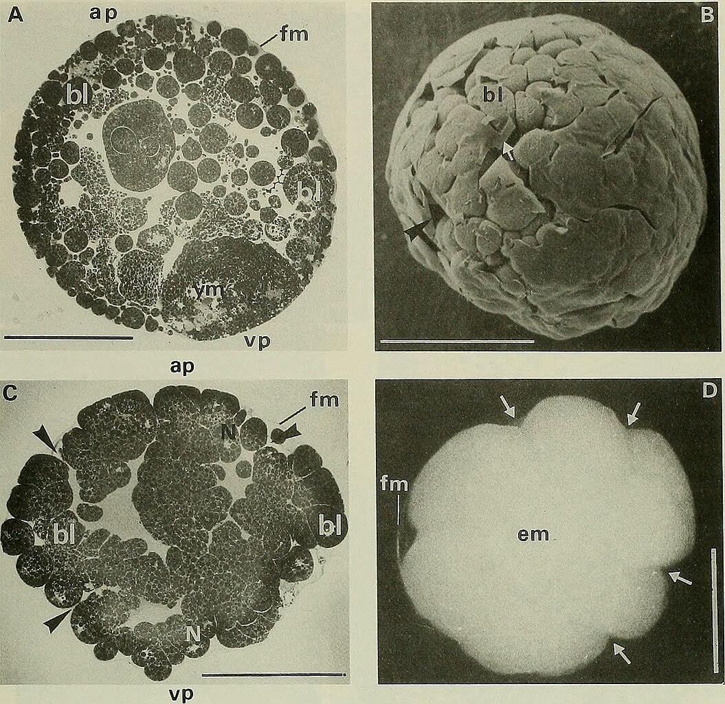
Figure 4. Segmentation of Abatus cordatus. (A) Section through a cleaved egg showing blastomeres and the remaining yolk mass at the vegetal pole (11 days after fertilization). (B) SEM view of a completely cleaved egg (14 days after fertilization). Blastomeres are visible where the fertilization membrane is destroyed (white arrow). Black arrow shows a furrow. (C) Section through a cleaved egg at the end of the segmentation (15 days after fertilization). Black arrows show the large intercellular spaces that communicate with the perivitelline space at the level of the furrows. (D) Light micrograph of a wrinkled stereoblastula (26 days after fertilization). Arrows show furrows. A and C are semi-thin plastic sections (toluidine blue, pH 11.5). Abbreviations: ap, animal pole; bl, blastomere; em, embryo; fm, fertilization membrane; N, nuclear area; vp, vegetal pole; ym, yolk mass. Scale bar = 500 um |
| Date | |
| Source | https://www.flickr.com/photos/internetarchivebookimages/20191999800/ The Biological bulletin. Marine Biological Laboratory (Woods Hole, Mass. ). Annual report; HighWire Press. |
| Author | Lillie, Frank Rattray, Moore, Carl Richard, Redfield, Alfred Clarence. |
Licensing[edit]
| This file is made available under the Creative Commons CC0 1.0 Universal Public Domain Dedication. | |
| The person who associated a work with this deed has dedicated the work to the public domain by waiving all of their rights to the work worldwide under copyright law, including all related and neighboring rights, to the extent allowed by law. You can copy, modify, distribute and perform the work, even for commercial purposes, all without asking permission.
|
| This image was originally posted to Flickr by Internet Archive Book Images at https://flickr.com/photos/126377022@N07/20191999800. It was reviewed on 25 March 2024 by FlickreviewR 2 and was confirmed to be licensed under the terms of the cc-zero. |
File history
Click on a date/time to view the file as it appeared at that time.
| Date/Time | Thumbnail | Dimensions | User | Comment | |
|---|---|---|---|---|---|
| current | 11:36, 25 March 2024 | | 1,640 × 1,592 (665 KB) | Rasbak (talk | contribs) | {{information |description= Figure 4. Segmentation of Abatus cordatus. (A) Section through a cleaved egg showing blastomeres and the remaining yolk mass at the vegetal pole (11 days after fertilization). (B) SEM view of a completely cleaved egg (14 days after fertilization). Blastomeres are visible where the fertilization membrane is destroyed (white arrow). Black arrow shows a furrow. (C) Section through a cleaved egg at the end of the segmentation (15 days after fertilization). Black arrows... |
You cannot overwrite this file.
File usage on Commons
The following 3 pages use this file:
File usage on other wikis
The following other wikis use this file:
- Usage on nl.wikipedia.org