File:10.1371 journal.pbio.0030137.g001-L-A.jpg
Jump to navigation
Jump to search
Size of this preview: 800 × 582 pixels. Other resolutions: 320 × 233 pixels | 640 × 465 pixels | 1,024 × 745 pixels | 1,280 × 931 pixels | 2,020 × 1,469 pixels.
Original file (2,020 × 1,469 pixels, file size: 514 KB, MIME type: image/jpeg)
Captions
Captions
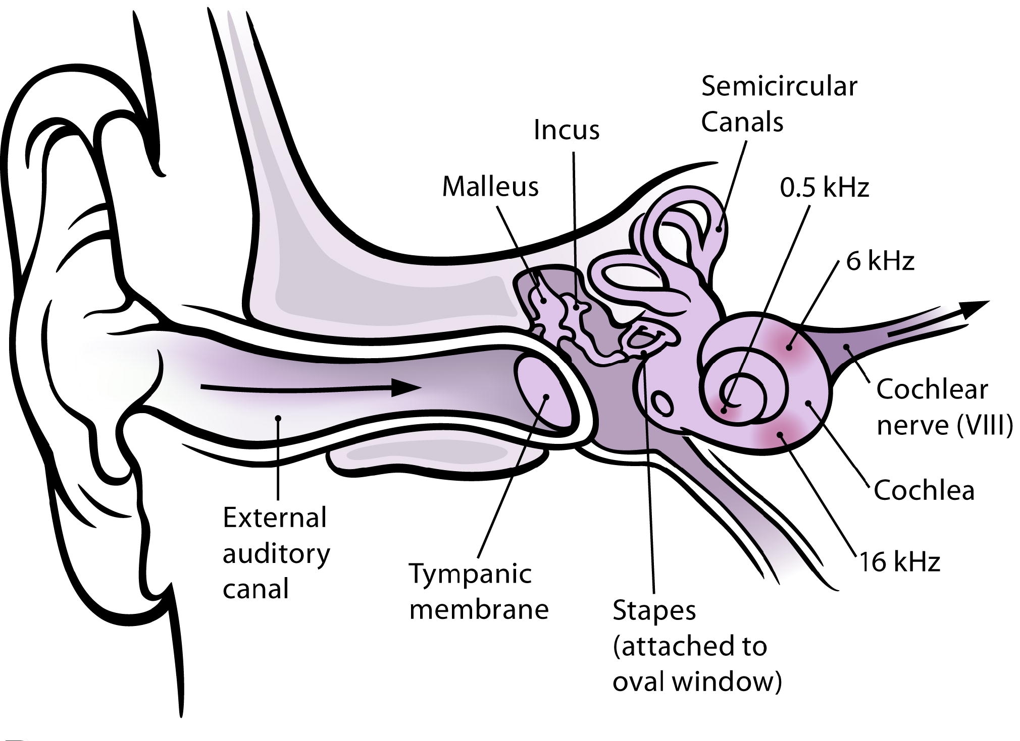
A labelled cross-sectional diagram of the human ear.

|
File:Anatomy of Human Ear with Cochlear Frequency Mapping.svg is a vector version of this file. It should be used in place of this JPG file when not inferior.
File:10.1371 journal.pbio.0030137.g001-L-A.jpg → File:Anatomy of Human Ear with Cochlear Frequency Mapping.svg
For more information, see Help:SVG. |
 |
| Description |
English: The human ear and frequency mapping in the cochlea. The three ossicles incus, malleus, and stapes transmit airborne vibration from the tympanic membrane to the oval window at the base of the cochlea. Because of the mechanical properties of the basilar membrane within the snail-shaped cochlea, high frequencies will produce a vibration peak near the oval window, whereas low frequencies will stimulate receptors near the apex of the cochlea (locations for three frequencies indicated schematically). Information from the cochlear receptor cells is transmitted to the cochlear nuclei via the 8th cranial nerve, and on through the midbrain to the cortex. (Redrawn from Figure 12.3 in [11].) |
| Date |
|
| Source | |
| Author |
|
| This is a retouched picture, which means that it has been digitally altered from its original version. Modifications: Isolated subfigure A. The original can be viewed here: 10.1371 journal.pbio.0030137.g001-L.jpg:
|
I, the copyright holder of this work, hereby publish it under the following licenses:
This file is licensed under the Creative Commons Attribution 2.5 Generic license.
- You are free:
- to share – to copy, distribute and transmit the work
- to remix – to adapt the work
- Under the following conditions:
- attribution – You must give appropriate credit, provide a link to the license, and indicate if changes were made. You may do so in any reasonable manner, but not in any way that suggests the licensor endorses you or your use.
You may select the license of your choice.
Original upload log[edit]
This image is a derivative work of the following images:
- File:10.1371_journal.pbio.0030137.g001-L.jpg licensed with Cc-by-2.5, Cc-by-2.5
- 2009-02-12T04:06:25Z Mike.lifeguard 2020x2480 (483539 Bytes) {{Information |Description={{en|1=(A) The human ear and frequency mapping in the cochlea. The three ossicles incus, malleus, and stapes transmit airborne vibration from the tympanic membrane to the oval window at the base of
Uploaded with derivativeFX
File history
Click on a date/time to view the file as it appeared at that time.
| Date/Time | Thumbnail | Dimensions | User | Comment | |
|---|---|---|---|---|---|
| current | 21:57, 28 April 2009 | | 2,020 × 1,469 (514 KB) | Mike.lifeguard (talk | contribs) | malleus and incus were swapped |
| 04:11, 12 February 2009 |  | 2,020 × 1,469 (521 KB) | Mike.lifeguard (talk | contribs) | {{Information |Description={{en|1=The human ear and frequency mapping in the cochlea. The three ossicles incus, malleus, and stapes transmit airborne vibration from the tympanic membrane to the oval window at the base of the cochlea. Because of the mechan |
You cannot overwrite this file.
File usage on Commons
The following 26 pages use this file:
- File:Anatomy of Human Ear with Cochlear Frequency Mapping.svg
- File:Anatomy of the Human Ear-Number.svg
- File:Anatomy of the Human Ear.svg
- File:Anatomy of the Human Ear 1 CAT.svg
- File:Anatomy of the Human Ear 1 Intl.svg
- File:Anatomy of the Human Ear ar.svg
- File:Anatomy of the Human Ear as.svg
- File:Anatomy of the Human Ear blank.svg
- File:Anatomy of the Human Ear cs-2.svg
- File:Anatomy of the Human Ear cs.svg
- File:Anatomy of the Human Ear id.svg
- File:Anatomy of the Human Ear in farsi numbers-edit-1.svg
- File:Anatomy of the Human Ear in farsi numbers.svg
- File:Anatomy of the Human Ear it.svg
- File:Anatomy of the Human Ear ja.svg
- File:Anatomy of the Human Ear kr.svg
- File:Anatomy of the Human Ear ku.svg
- File:Anatomy of the Human Ear mk.svg
- File:Anatomy of the Human Ear nan.svg
- File:Anatomy of the Human Ear no.svg
- File:Anatomy of the Human Ear pl.svg
- File:Anatomy of the Human Ear pt.svg
- File:Anatomy of the Human Ear ro.svg
- File:Anatomy of the Human Ear sv.svg
- File:Anatomy of the Human Ear uk.svg
- Template:Other versions/Anatomy of the Human Ear
File usage on other wikis
The following other wikis use this file:
- Usage on eu.wikipedia.org
- Usage on sr.wikipedia.org
- Usage on zh-yue.wikipedia.org