Commons:Featured picture candidates/File:Eye orbit anatomy anterior2.jpg
Jump to navigation
Jump to search

File:Eye orbit anatomy anterior2.jpg, featured[edit]
Voting period is over. Please don't add any new votes.Voting period ends on 24 Jul 2011 at 14:40:08 (UTC)
Visit the nomination page to add or modify image notes.

 Info created & uploaded by Patrick.lynch - nominated by Tomer T (talk) 14:40, 15 July 2011 (UTC)
 Support -- Tomer T (talk) 14:40, 15 July 2011 (UTC)
- Support -- Raghith 10:56, 16 July 2011 (UTC)
 Question As I'm nor an anatomist neitehr an ophtalmologist, I have a question: is this picture complete ? Is there nothing else than eye and his muscles to be shown only, is it normal for the bone of the skull to be "naked" like this ? I'm not sure about the full educational value of this design... Moreover, the blue background is noisy...--Jebulon (talk) 17:06, 16 July 2011 (UTC)
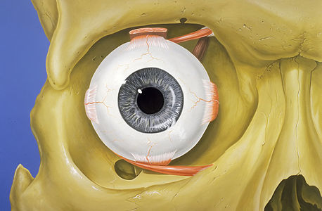
 Comment You're right, for sure, regularly around the eye there are many tissues (as you know there are epithelial tissue, connective tissue, muscle tissue and nervous tissue; specifically for this presentation we need muscle and nervous tissue, to highlight the main point — the eye and its connection to brain.) The author has wanted to present only the anatomy of the eye (the sclera, cornea, retina, pupil... ...and nervous connection of the eye and brain, that's the optic nerve and retinal blood vessels) — we need only muscle and nervous tissue here, so showing complete facial musculature around the eye would be pointless for educative experience of the picture. I hope u're getting this. Alex discussion 22:03, 16 July 2011 (UTC)
 Neutral I will support it if it is denoised,
- Support As I am 2nd class student at High Medical School, I can very precisely figure out what's good on this photo. It is about a very well and creative illustrated image of human eye anatomy. Also, it's kind of remarkable resolution and sharpness, but that noise could be fixed in alternative version. Anyway, this is amazing and I'll support it in advance. Alex discussion 21:33, 16 July 2011 (UTC)
- Support For educational value regarding human anatomy. The noise in the blue background can easily be cleaned out. —stay (sic)! 00:45, 17 July 2011 (UTC)
 Noise is gone. W.S. 07:25, 20 July 2011 (UTC)
- Support --Jovian Eye talk 10:14, 20 July 2011 (UTC)
- Support --Karelj (talk) 20:53, 21 July 2011 (UTC)
- Support Very good now! -- H005 21:53, 21 July 2011 (UTC)
- Support Miam--Citron (talk) 17:08, 23 July 2011 (UTC)
Confirmed results:
Result: 8 support, 0 oppose, 0 neutral → featured. /George Chernilevsky talk 17:39, 24 July 2011 (UTC)
This image will be added to the FP gallery: Non-photographic media/Computer-generated